About us — the people behind GO Privatpraxis
A team that takes its time
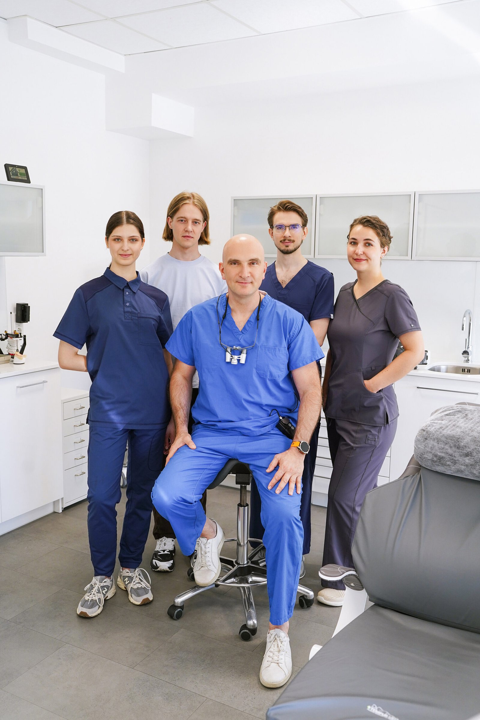
Five people, one dentist per patient and the microscope at every step. Here the treatment is not the focus, the person is — which is why we schedule appointments so that there is time for questions.
Our approach
The machine does not heal. The dentist does.
Modern equipment, digital diagnostics, high-end technology — we have all of it. But technology is a tool, not a promise. What matters is who guides it.
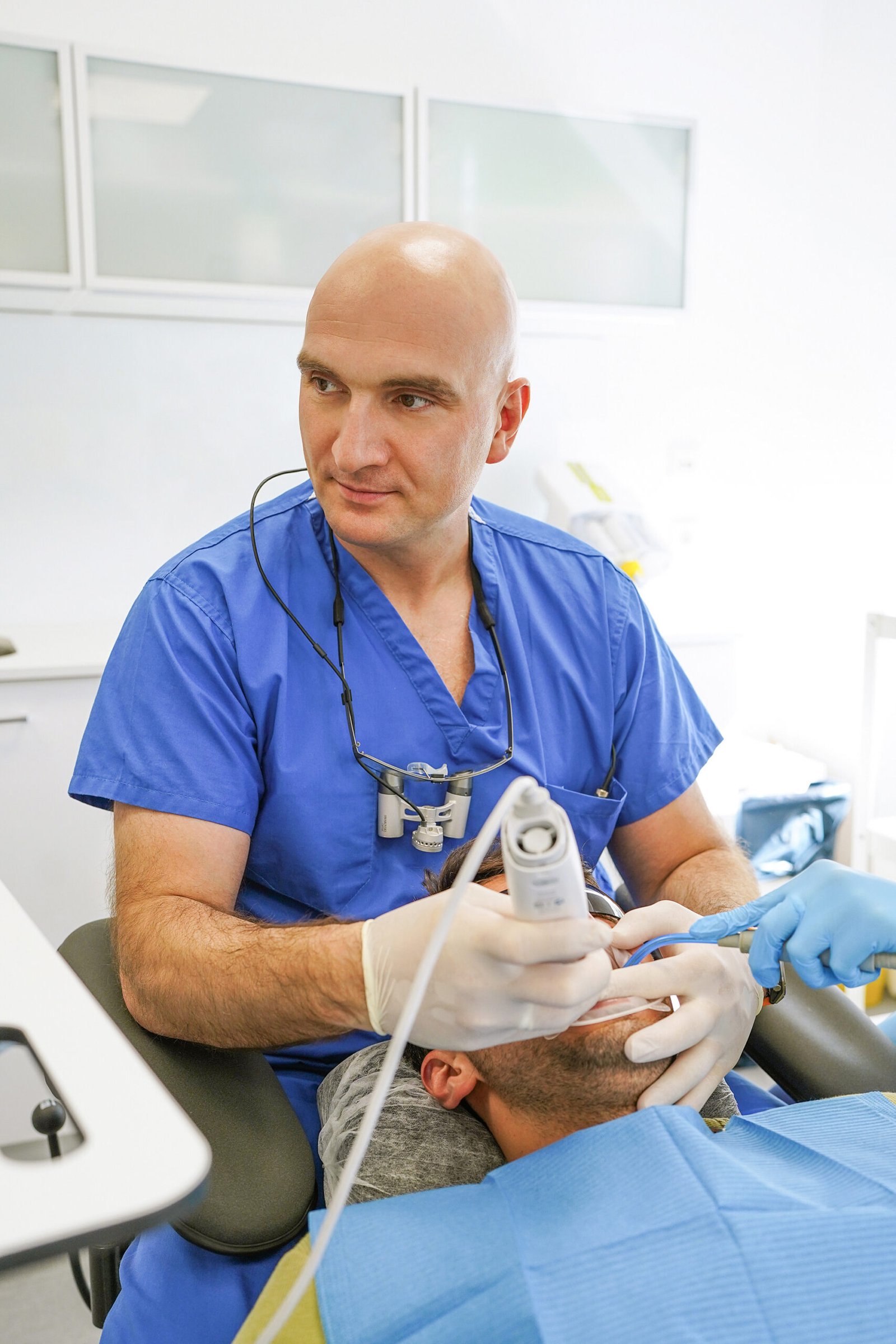
That is why one dentist accompanies you from the first examination to the final check-up. No change of hands, no handover, no telling your story a second time.
“I treat the way I would want to be treated myself: with time, with explanation and under the microscope — even when it would be quicker without.”Dr Givi Ordzhonikidze, practice ownerOur treatments →
The team
Five who work with each other, not alongside each other
Three dentists, a specialist for the digital treatment workflow and an assistant who knows every move. Short distances within the team mean short distances for you.
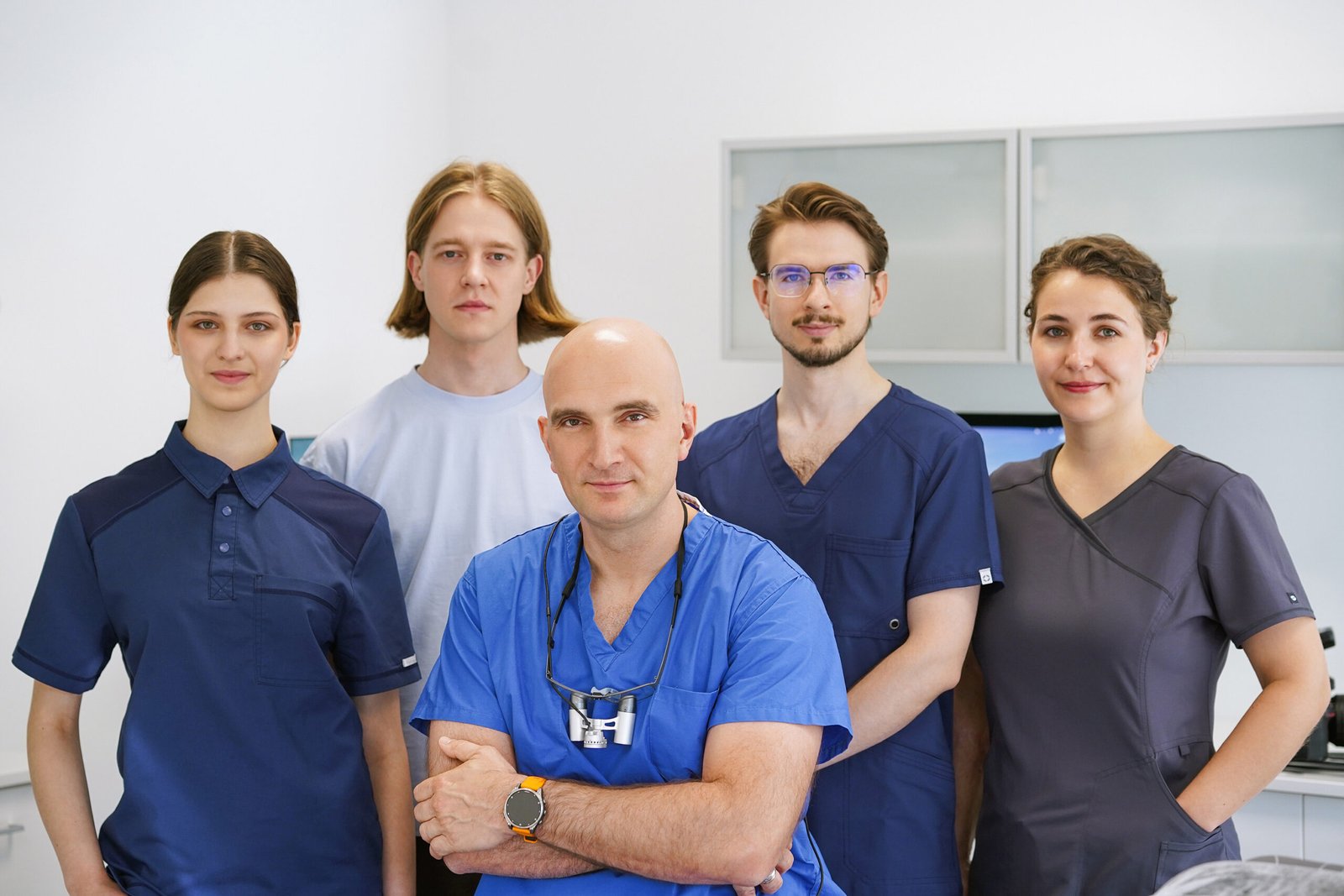
- 01Dr Givi OrdzhonikidzeDentist · practice owner · functional diagnostics and implantology
- 02Dr Dmitri SolomatnikovDentist
- 03Dr Anastasia SolomatnikovaDentist
- 04Miroslav MalchukSpecialist for the digital treatment workflow — scanning, planning, fabrication
- 05Anastasia TichonowaDental assistant — assisting Dr Ordzhonikidze
Particular expertise
When the pain does not come from the tooth
Our strength — and almost unique in Germany and the Netherlands — is the use of the Gamma Condylograph. The device records the movements of your jaw joint in three dimensions and makes visible what an X-ray cannot show.
This allows us to help patients who have long searched elsewhere for answers without success.
- Jaw joint problems — clicking, grinding, restricted mouth opening
- Neck and back complaints with a possible cause in the bite
- Chronic headaches and muscle tension
- Migraine, where dental causes are a possibility
How we work
Five steps that are part of every treatment here
Not a marketing promise, but the process you will actually experience with us.
Findings, images and consultation
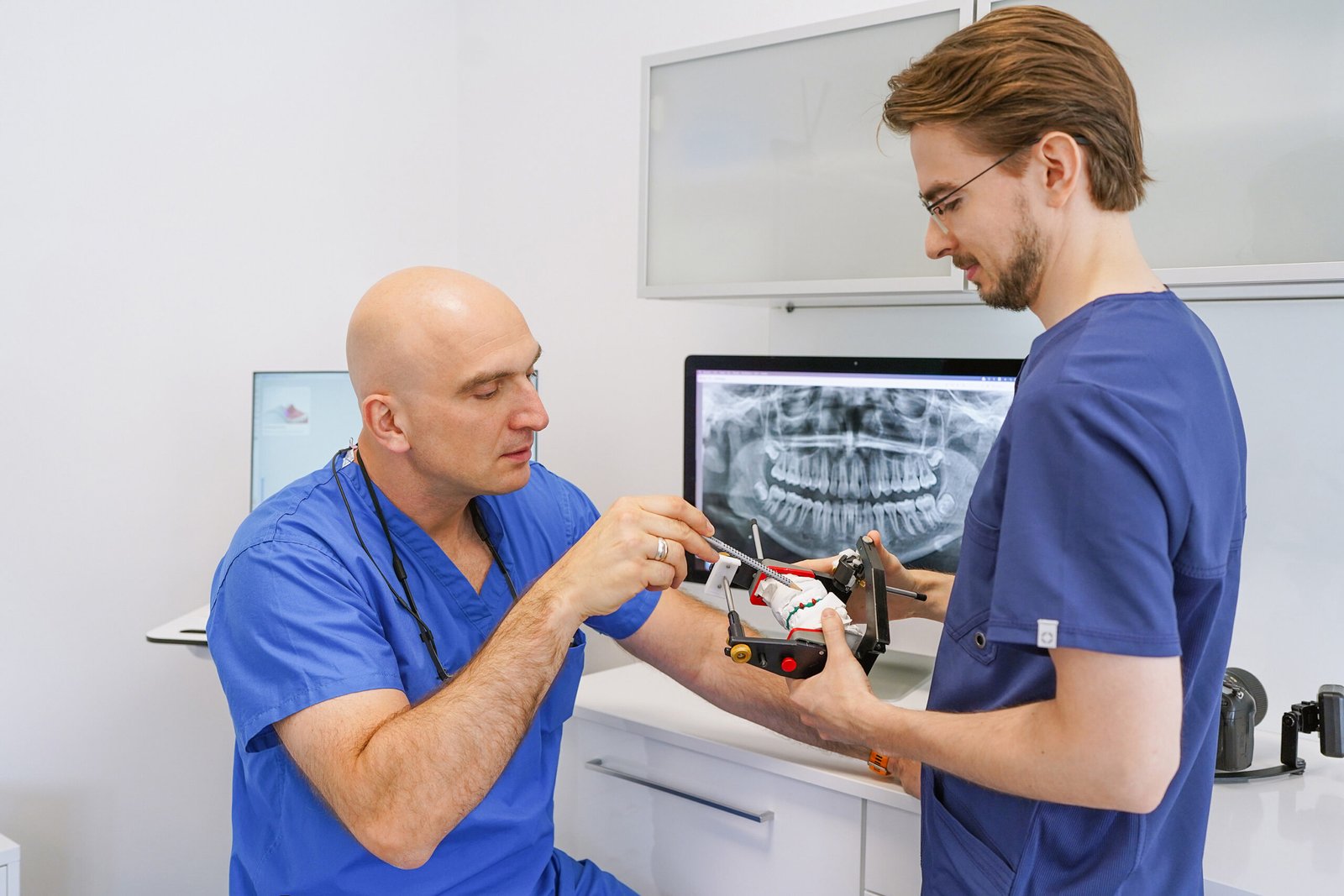
At the beginning there is no treatment plan, but a picture: X-ray, photo status, functional analysis. We show you on the monitor what we see and explain why we recommend what we recommend — and what the alternatives are.
Treatment under the microscope
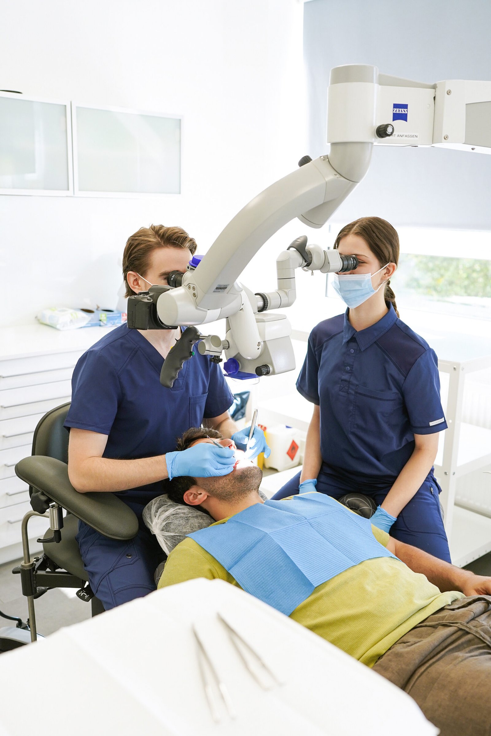
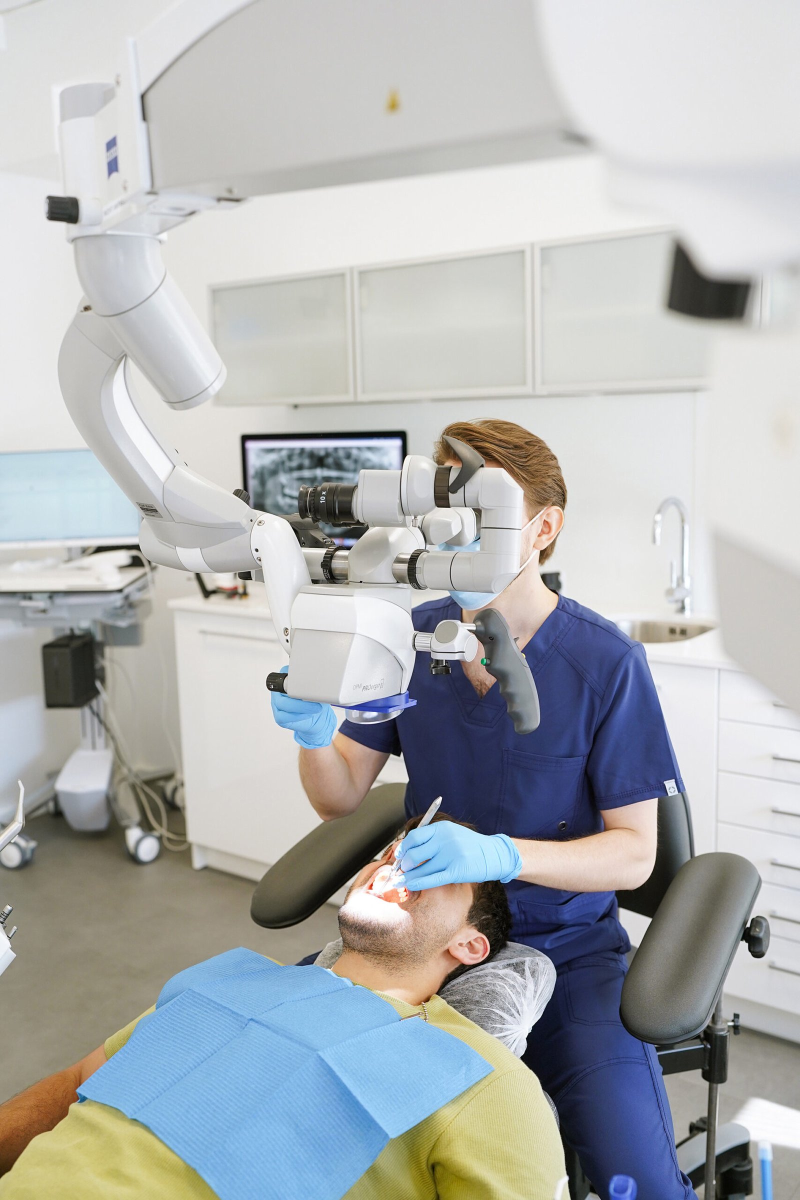
What the eye cannot see cannot be treated cleanly. We work with the ZEISS operating microscope — for root canal treatment just as for preparing a crown. The result is smaller accesses, more precise margins and less rework.
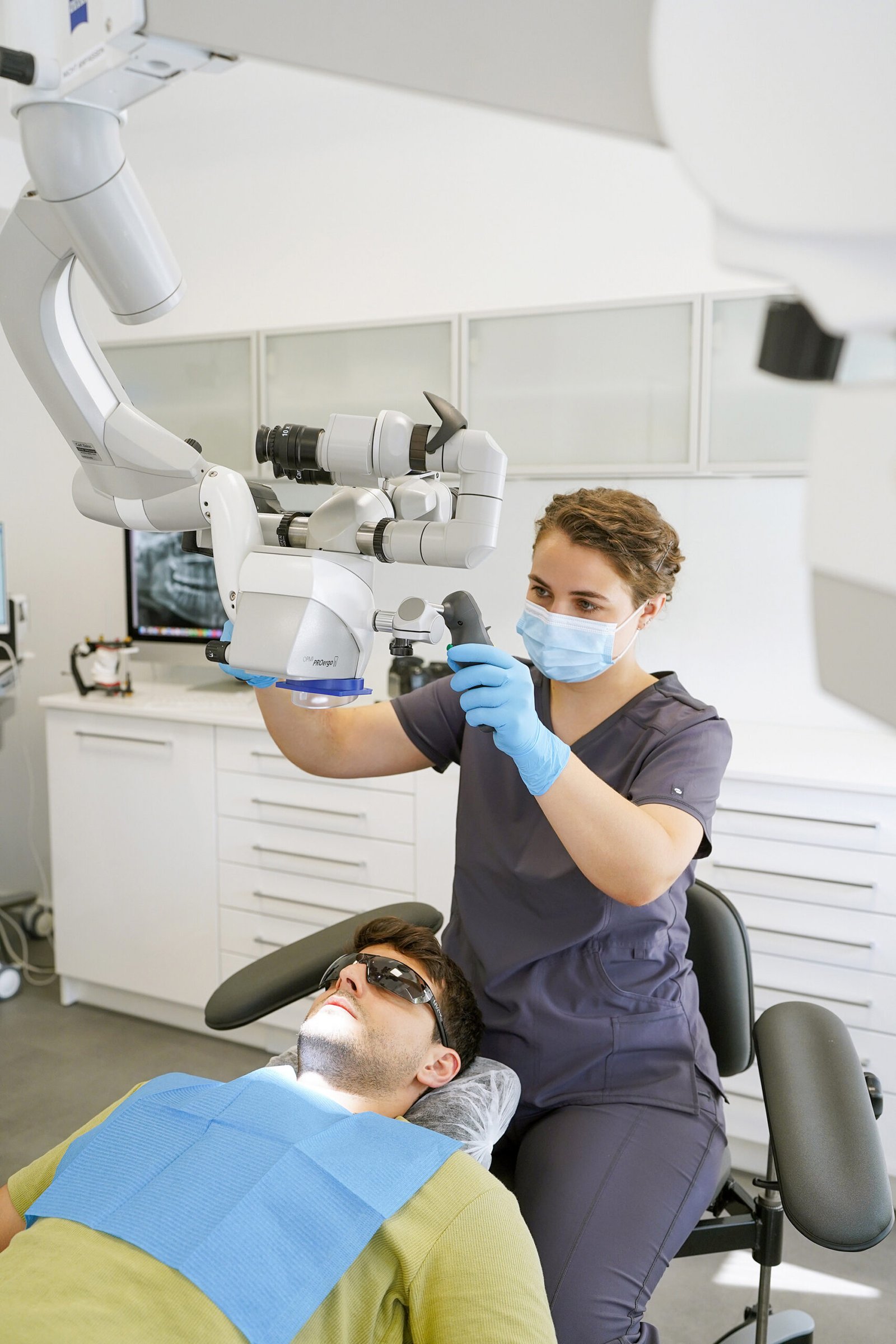
Four hands, one focus
Every treatment is assisted. That shortens your time in the chair and keeps the dentist where they belong: concentrating on the tooth rather than on the instrument tray.
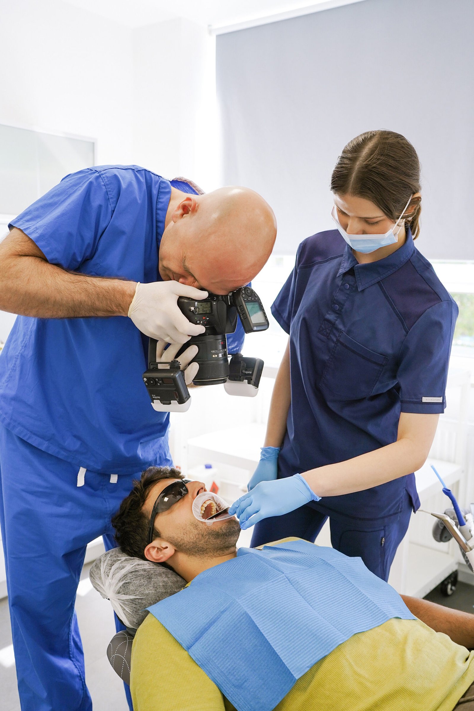
Every step is documented
Starting point, intermediate steps, result: we photograph as we go. For you as traceable evidence, for us as quality control — and as a basis if a question comes up years later.
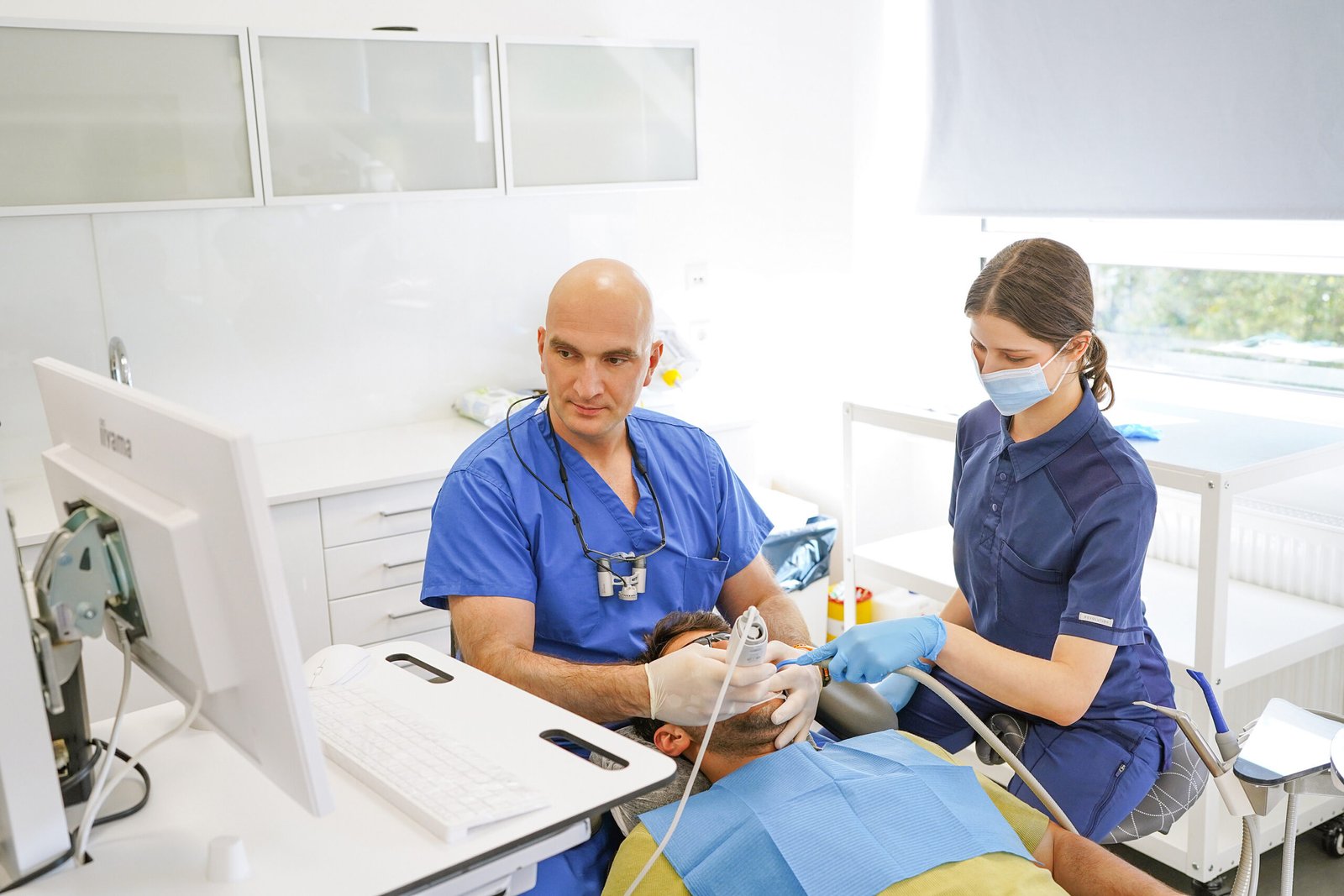
Planned digitally, made in-house
Digital impression, planning on screen, fabrication in our own Dent-Tech milling centre in Wuppertal. Short distances mean short waiting times — and corrections that can often be made the same day.
Technology
Equipment you can see in the pictures
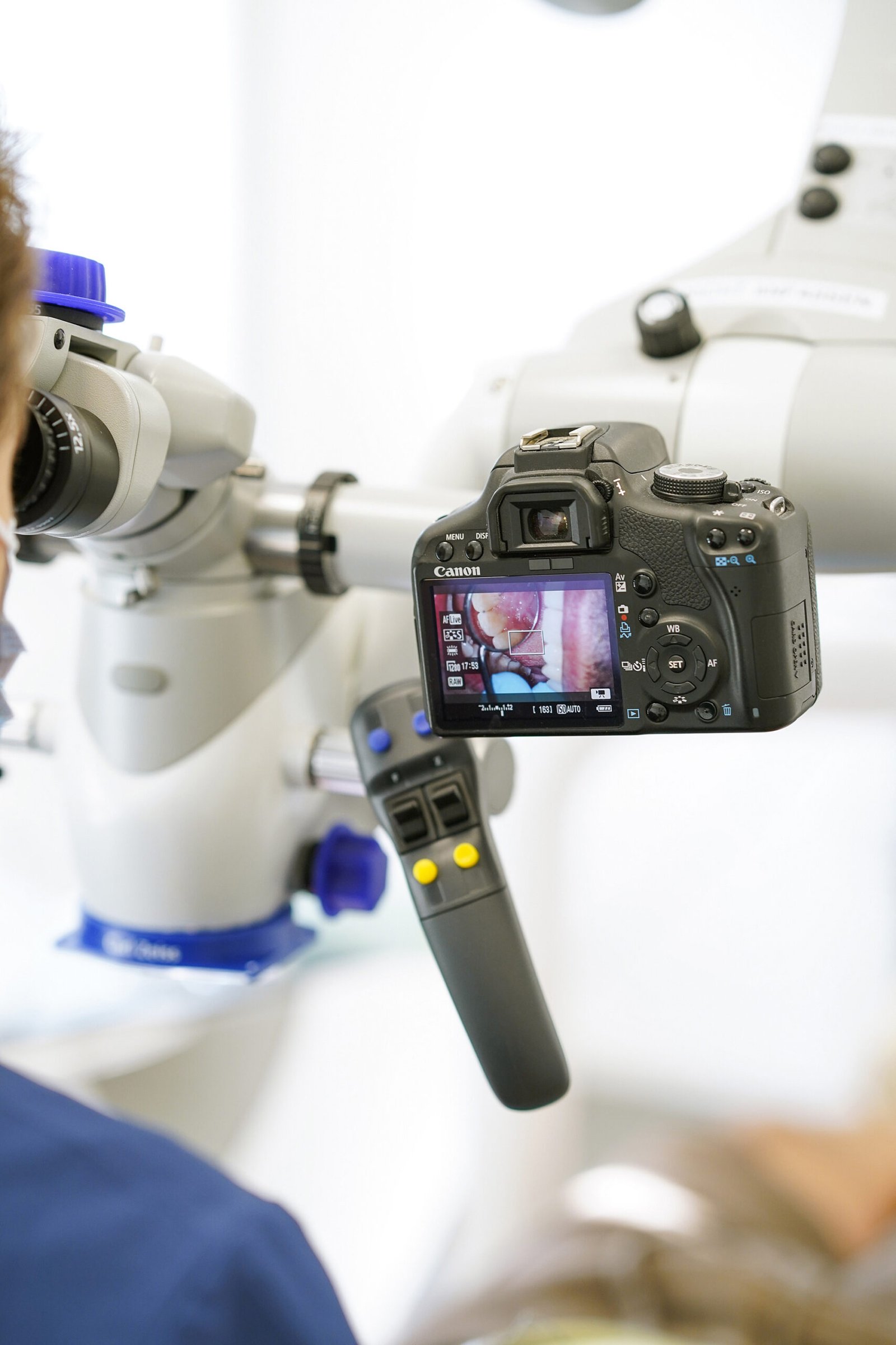
ZEISS operating microscope
With an attached camera — the same optics that treat also document. Afterwards you see what we saw.
Digital X-ray at the chair
Images are available at the treatment station within seconds — low radiation dose, no waiting, discussed with you directly.

Our own milling centre
Dental restorations are made at Dent-Tech in the same building. We talk to the technician down the corridor — instead of emailing a laboratory in another city.